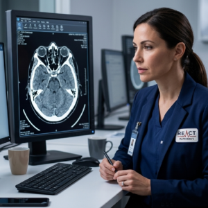
Crucial for frontline healthcare professionals managing acute neurological presentations or trauma:
 Advanced Clinical Practitioners (ACPs) and ACP TraineesMedical Students and Junior DoctorsNurses and Allied Health Professionals working in acute neuro or trauma environmentsEmergency Medicine, Neurology, and Trauma CliniciansHealthcare Professionals working in hospital and urgent care unitsFlexible E-Learning: Self-paced, remote digital learning featuring interactive clinical cases and step-by-step guided explanations.Face-to-Face Teaching: Dynamic, classroom-based workshops packed with facilitated peer discussions, practical case applications, and expert clinician-led teaching.
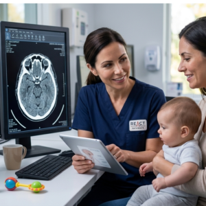 Anatomical Review Frameworks: Structured systems to systematically evaluate the entire intracranial landscape.Emergency Haemorrhage Identification: Deep dives into distinct, high-risk intracranial bleeding patterns.Ischaemic vs. Haemorrhagic Contrast: Master the key imaging features that dictate contrasting stroke pathways.Escalation Logic: Clear guidelines on when to urgently escalate to specialist review or advanced MRI imaging.
By the end of this module, you will be able to:
Apply a rigorous, structured approach to evaluate brain parenchyma, ventricles, midline shifts, and extra-axial spaces.Differentiate instantly between subdural, extradural, subarachnoid, and intracerebral haemorrhages.Spot early radiographic signs of acute ischaemic stroke, cerebral oedema, and mass effect.Identify subtle skull fractures and structural abnormalities.Safely correlate CT head findings with your patient's physical neurological examination and clinical history.